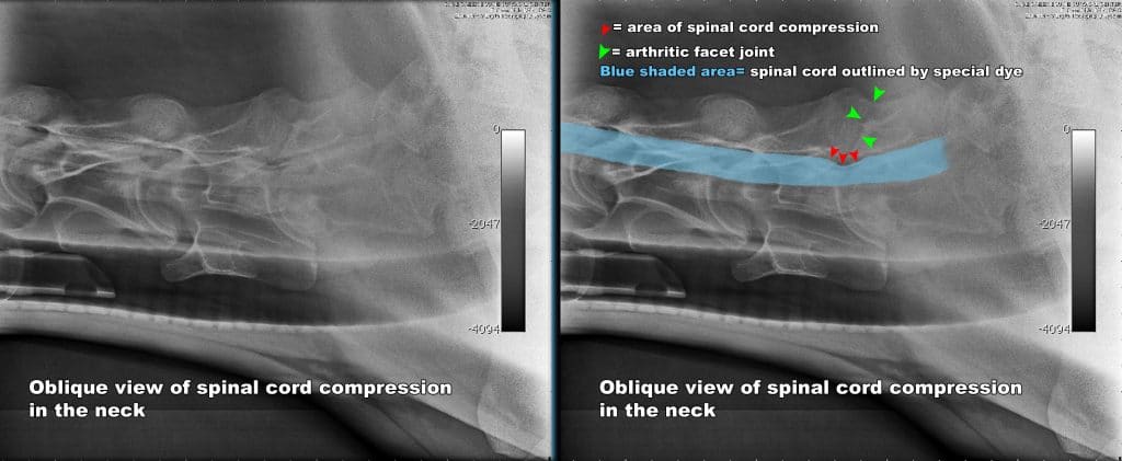
An equine wobbler is a horse with a damaged spinal cord. This can occur from malformation of the vertebral column, advanced arthritis in the vertebral joints or injury to the vertebrae. Any of these conditions can cause pressure on the spinal cord and cause the flow of information between the limbs and the brain to become obstructed.
A horse with wobblers may stumble, wear his toes abnormally, over-reach and clip the heels of the forelimbs, ‘bunny hop’ when cantering, or show excessive knuckling of hind legs. Most horses with this condition show more pronounced signs in the hindlimbs. This is because the nerves that go to/from the hindlimbs travel down the outside of the spinal cord, so are prone to injury. The nerves to/from the forelimbs are protected deeper within the spinal cord.
When a horse shows signs of spinal cord damage we say he is neurologic. There are 5 neurologic grades:
- Grade 1- Very slight signs
- Grade 2- Clumsy gait
- Grade 3- Looks drunk
- Grade 4- Falling down
- Grade 5- Can’t stand up
Diagnosis
With low grade deficits, it is easy to mistake a neurologic problem for a lameness problem. Many owners just think their horse is clumsy or needs to get in better condition. It takes a detailed neurologic exam, performed by an experienced clinician to detect a mild neurologic deficit.
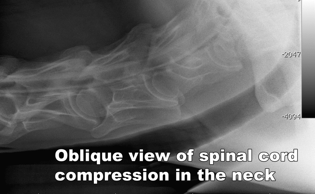
Once a horse is determined to have a neurologic problem, then diagnostic tests can be performed. X-rays and myelogram are the best ways to see the bones and spinal cord in neck area (where most wobbler conditions occur). X-rays can show fractures (new and old), arthritis, and areas of instability.
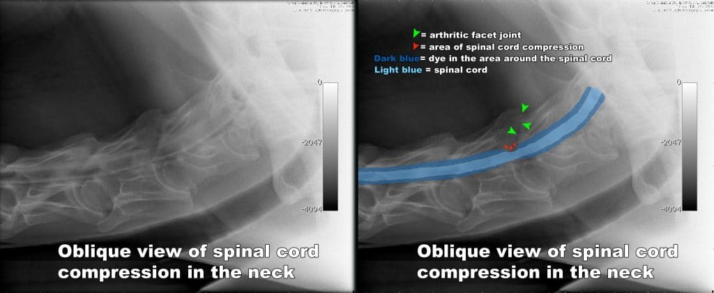
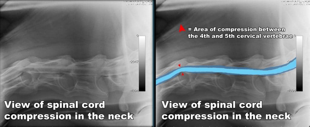
A myelogram is performed under IV anesthesia to check compression of the spinal cord. An X-ray opaque dye is injected into the small space around the spinal cord, then a series of X-rays is taken. Normally the spinal cord is invisible to X-rays, but when outlined by the dye, its borders can be clearly seen.
Below are images of a myelogram showing compression of the spinal cord from arthritis in the dorsal facet joint between the 6th and 7th cervical vertebrae and from instability between the 4th and 5th cervical vertebrae.
Treatment
Surgical fusion of the affected spinal vertebrae is indicated to help alleviate pressure on the spinal cord. The horse is placed under general anesthesia on his back. The neck is extended, and special stainless steel ‘baskets’ are screwed into place in the area of the vertebral disc. The baskets are filled with the bone fragments that are removed during the drilling process. With time, the bone fuses through the basket, and the vertebrae are permanently fixed in place.

Positioning the patient for surgery. The surgical site is cleaned and x-rays are taken to localize the area to be treated.

Once the patient is draped in sterile drapes, the surgery is performed using specialized tools specifically designed for this procedure. Dr. Barry Grant is performing this procedure with Dr. Elaine Carpenter assisting.
Post operative x-ray showing the baskets in place. The bone will fuse thru the baskets and the vertebrae will be fixed in place.
Prognosis
Typically 80% of all patients can improve at least one grade and 54% can improve two grades or more with surgery. Thirty-three percent of horses perform athletically. Horses should be graded <1/5 before beginning athletic work.

